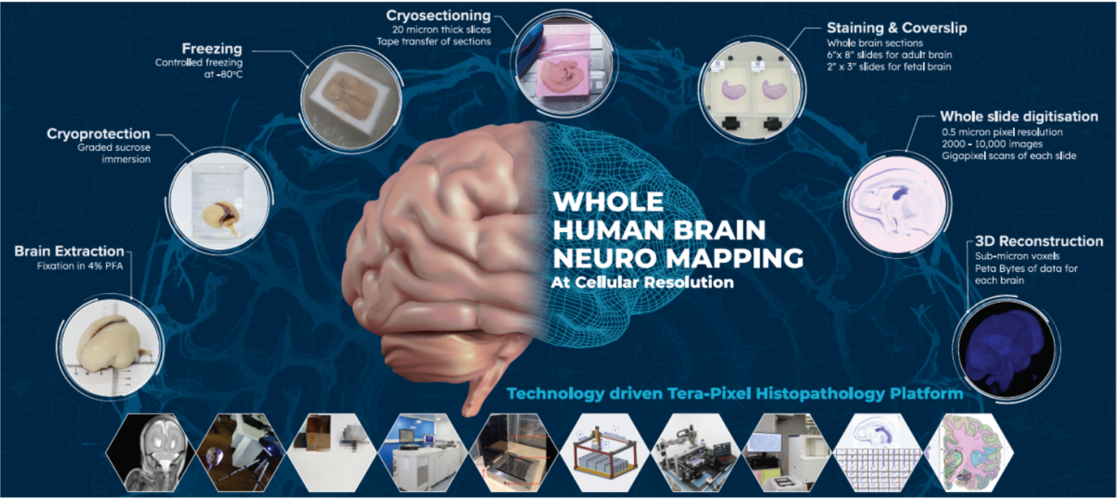
Technology platform for freezing large brains in stereotaxic coordinates with minimal tissue damage or artifacts.
Automated system for application of 10-20μm thin adhesive layer on glass slides to preserve tissue integrity during tape transfer cryo-sectioning

An automated system for cryo-sectioning and transfer of tissue to glass slides with minimal tissue damage.
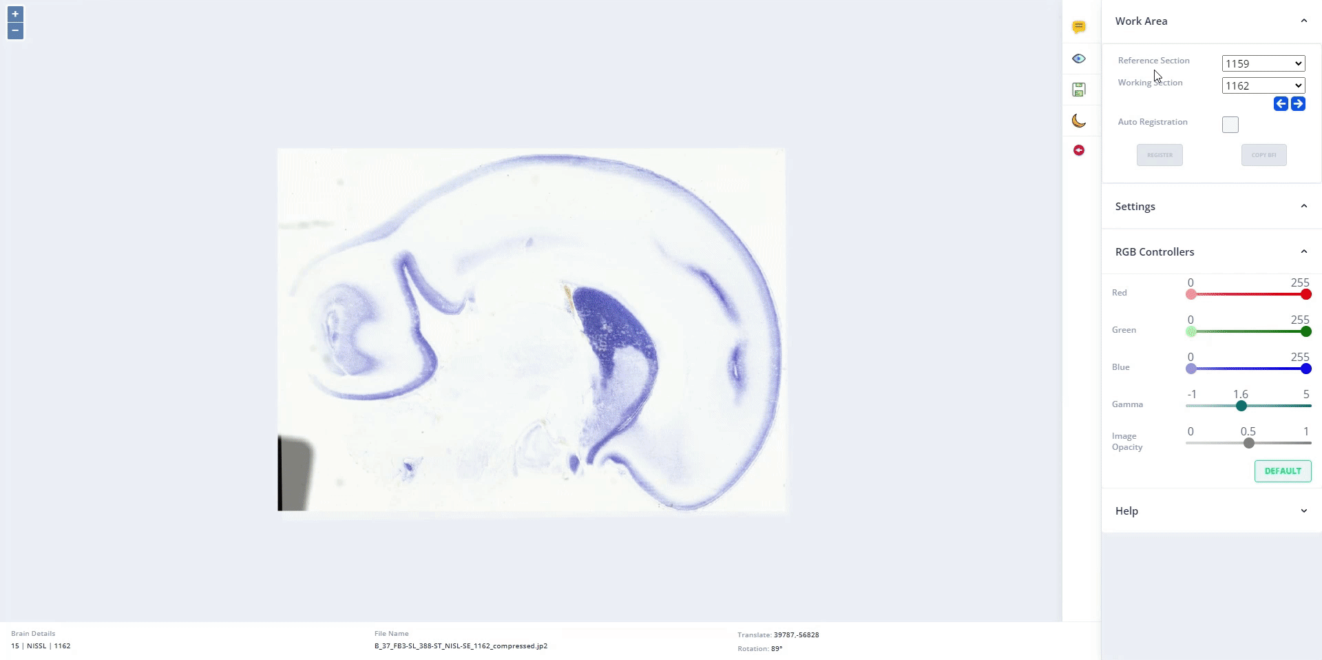
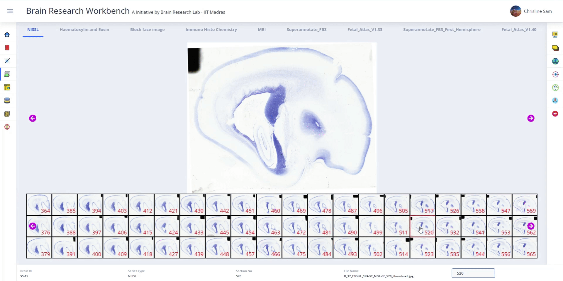
An intermediate step in histopathology pipeline to detect structure and volume changes of the brain using imaging.
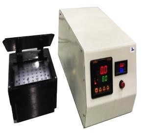
Hands-free, tool-free method of transferring a 20-micron thick sectioned tissue onto a glass slide via controlled UV exposure
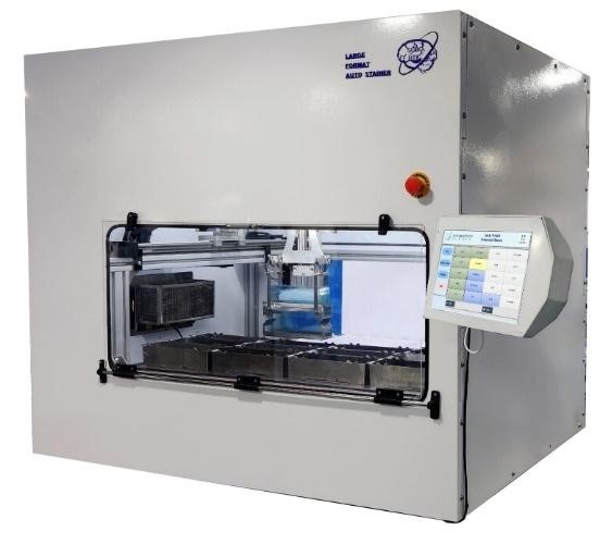
A custom-built automated staining system for tissues mounted on multiformat slides (6x8, 5x7, 2x3, and 1x3) with uniformity and consistency.
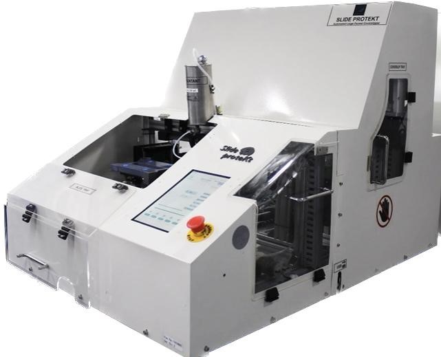
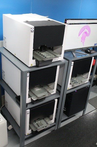
An automated system for uniform, bubble-free cover slipping operation of (6x8)slides with a throughput of 30 slides/hour.
A fully automated system capable of cover slipping (2x3) slides with a high throughput of 50 slides/hour.
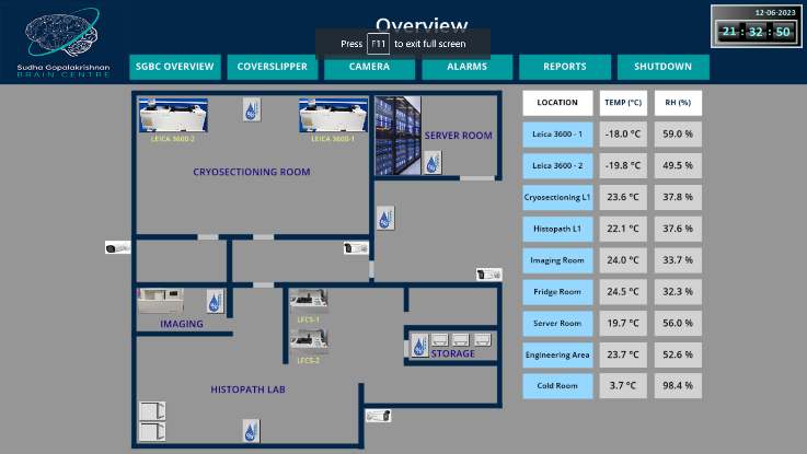
Monitors critical operations in the lab - environmental parameters (temperature, humidity), coverslipping & staining. Generates reports and alerts users during failure
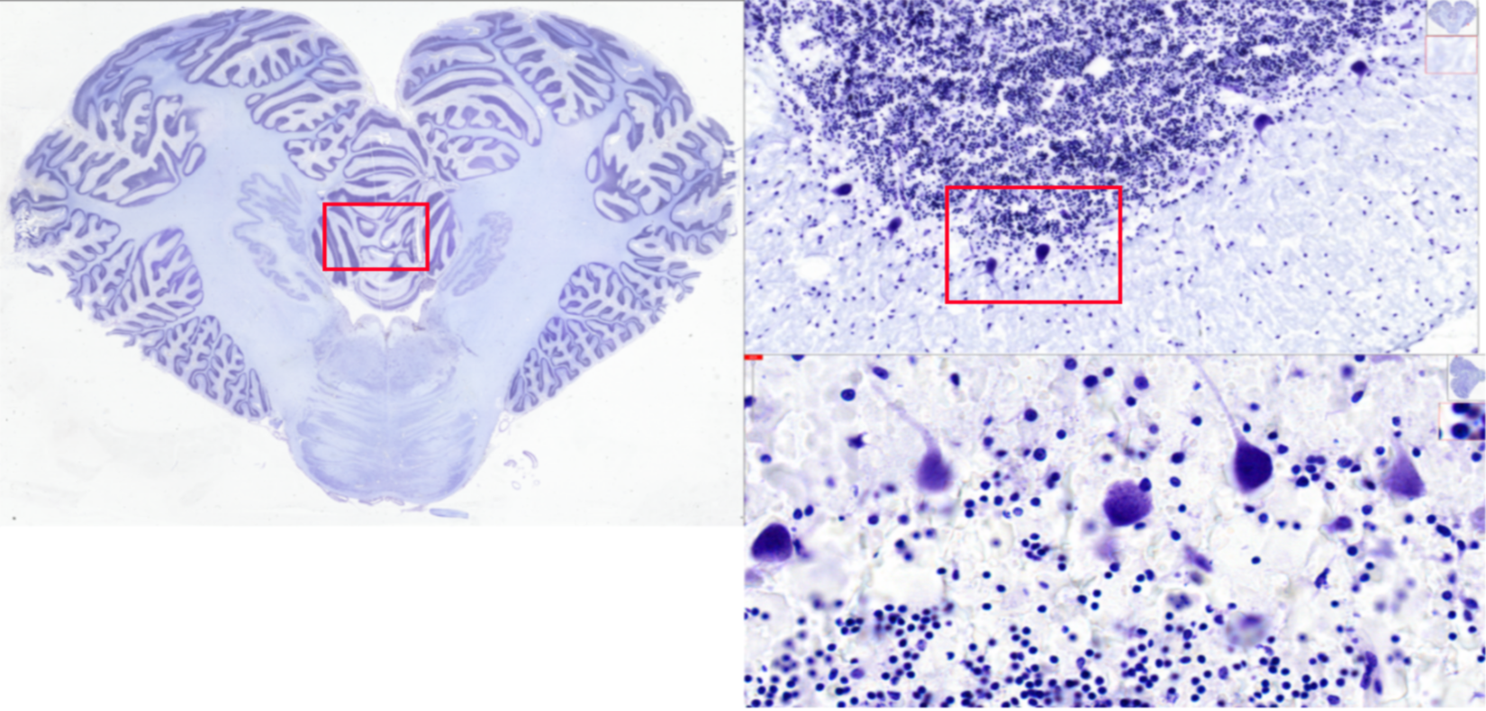
Custom whole slide scanners for digitizing multi format slides at 0.5 μm/pixel resolution - developed in collaboration with an indigenous company.
DHARANI: A 3D Developing Human-Brain Atlas Resource to Advance Neuroscience Internationally Integrated Multimodal Imaging and High-Resolution Histology of the Second Trimester
Richa Verma, Mihail Bota, Keerthi Ram, Jaikishan Jayakumar, Rebecca Folkerth, Karthika Pandurangan, Jivitha Jyothi Ramesh, Moitrayee Majumder, Rakshika Raveendran, Reetuparna Nanda, Sivamani K, Amal Dhivahar S, Srinivasa Karthik, Ramdayalan Kumarasami, Suresh S, S. Lata, E. Harish Kumar, Rajeswaran Rangasami, Chitra Srinivasan, Jayaraman Kumutha, Sudha Vasudevan, Koushik Bhat, Chrisline Sam C, Sivathanu Neelakantan, Stephen Savoia, Partha P. Mitra, Jayaraj Joseph, Paul R. Manger, Mohanasankar Sivaprakasam
Journal of Comparative Neurology, Volume 533, Issue 2, 2025, e70006. doi: 10.1002/cne.70006
SHFormer: Dynamic spectral filtering convolutional neural network and high-pass kernel generation transformer for adaptive MRI reconstruction
Sriprabha Ramanarayanan, Rahul G.S., Mohammad Al Fahim, Keerthi Ram, Ramesh Venkatesan, Mohanasankar Sivaprakasam
Neural Networks, Volume 187, 2025, 107334. doi: 10.1016/j.neunet.2025.107334
KDPhys: An attention guided 3D to 2D knowledge distillation for real-time video-based physiological measurement
Nicky Nirlipta Sahoo, VS Sachidanand, Matcha Naga Gayathri, Balamurali Murugesan, Keerthi Ram, Jayaraj Joseph, Mohanasankar Sivaprakasam
Biomedical Signal Processing and Control, Volume 107, 2025, 107797. doi :10.1016/j.bspc.2025.107797
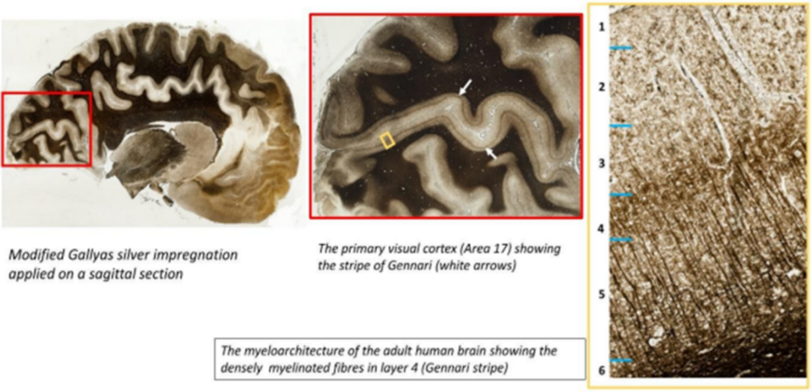
The histological development of the fetal human inferior colliculus during the second trimester
Nanda Reetuparna, Bota Mihail, Jayakumar Jaikishan, S Suresh, Lata S., Kumar E. Harish, Srinivasan Chitra, Vasudevan Sudha, Jayaraman Kumutha, Sivaprakasam Mohanasankar, Verma Richa
Frontiers in Neuroanatomy, Volume 18, 2025. doi: 10.3389/fnana.2024.1502778
AutoDPS: An unsupervised diffusion model based method for multiple degradation removal in MRI
Arunima Sarkar, Ayantika Das, Keerthi Ram, Sriprabha Ramanarayanan, Suresh Emmanuel Joel, Mohanasankar Sivaprakasam
Computer Methods and Programs in Biomedicine, Volume 263, 2025. doi: 10.1016/j.cmpb.2025.108684.
AAD-DCE: An Aggregated Multimodal Attention Mechanism for Early and Late Dynamic Contrast Enhanced Prostate MRI Synthesis
Divya Bharti, Sriprabha Ramanarayanan, Sadhana S, Kishore Kumar M, Keerthi Ram, Harsh Agarwal, Ramesh Venkatesan, Mohanasankar Sivaprakasam
IEEE International Conference on Acoustics, Speech, and Signal Processing (ICASSP 2025) doi: 10.48550/arXiv.2502.02555
Predictive modeling of Alzheimer's disease progression: Integrating temporal clinical factors and outcomes in time series forecasting
K.H. Aqil , Prashanth Dumpuri , Keerthi Ram, Mohanasankar Sivaprakasam
Intelligence-Based Medicine Volume 10, 2024, 100159 doi: 10.1016/j.ibmed.2024.100159
Systematic development of immunohistochemistry protocol for large cryosections-specific to non-perfused fetal brain
Karthika Pandurangan , Jaikishan Jayakumar , Stephen Savoia , Reetuparna Nanda , S. Lata , E. Harish Kumar , Suresh S. , Sudha Vasudevan , Chitra Srinivasan , Jayaraj Joseph, Mohanasankar Sivaprakasam, Richa Verma
J Neurosci Methods. 2024 May:405:110085. doi: 10.1016/j.jneumeth.2024.110085.
Histological characterization and development of mesial surface sulci in the human brain at 13–15 gestational weeks through high-resolution histology
Richa Verma, Jaikishan Jayakumar, Rebecca Folkerth, Paul R. Manger, Mihail Bota, Moitrayee Majumder, Karthika Pandurangan, Stephen Savoia, Srinivasa Karthik, Ramdayalan Kumarasami, Jayaraj Joseph, G. Rohini, Sudha Vasudevan, Chitra Srinivasan, S. Lata, E. Harish Kumar, Rajeswaran Rangasami, Jayaraman Kumutha, S. Suresh, Goran Šimić, Partha P Mitra, Mohanasankar Sivaprakasam
J Comp Neurol. 2024 Apr;532(4):e25612. doi: 10.1002/cne.25612.
Ano-swinMAE: Unsupervised Anomaly Detection in Brain MRI using swin Transformer based Masked Auto Encoder
Kumari Rashmi, Ayantika Das, Naga Gayathri Matcha, Keerthi Ram, Mohanasankar Sivaprakasam
MIDL 2024
Registration Quality Evaluation Metric with Self-Supervised Siamese Networks
Tanvi Kulkarni, Sriprabha Ramanarayanan, Keerthi Ram , Mohanasankar Sivaprakasam
MIDL 2024
Optimization of Freezing Method to Facilitate Cryosectioning of Large Brain Tissues
Ramdayalan Kumarasami, Sathish Pandidurai, Srinivasa Karthik, Mohanasankar Sivaprakasam, Jayaraj Joseph
2024 IEEE International Symposium on Medical Measurements and Applications (MeMeA) doi: 10.1109/MeMeA60663.2024.10596795
Advancing Neuroscience Research with Visual Question Answering and Multimodal Retrieval
Pralaypati Ta, Sriram Venkatesaperumal, Mohanasankar Sivaprakasam, Amit Kumar and Vega Shah
Technical Blog , nvidia.Developer
Automated Coverslipper for Large Format Slides with Switchable Compatibility to Handle Multi Format Slides.
Jayaraj Joseph, Mohanasankar Sivaprakasam, Jayaraman Kiruthi Vasan, Ramdayalan K, Rahul Manoj, and Sudhan Chandrasekaran
WO 2023/275894, Jan. 5, 2023, Assigned to Indian Institute of Technology (IIT Madras).
A Standardized Protocol for the Safe Retrieval of Infectious Postmortem Human Brain for Studying Whole-Brain Pathology
James RI, Verma R, Johnson LR, Manesh A, Jayakumar J, Sen M, Joseph J, Kumarasami R, Mitra PP, Sivaprakasam M, Varghese GM
Am J Forensic Med Pathol. 2023 Jul 25. doi: 10.1097/PAF.0000000000000871. Epub ahead of print. PMID: 37490584.
A technology platform for standardized cryoprotection and freezing of large-volume brain tissues for high-resolution histology
Ramdayalan Kumarasami , Richa Verma , Karthika Pandurangan , Jivitha Jyothi Ramesh, Sathish Pandidurai, Stephen Savoia, Jaikishan Jayakumar , Mihail Bota , Partha Mitra, Jayaraj Joseph , Mohanasankar Sivaprakasam
Front. Neuroanat., 02 November 2023 Volume 17 - 2023 doi: 10.3389/fnana.2023.1292655
Wide field block face imaging using deep ultraviolet induced autofluorescence of the human brain
Karthik, S., Joseph, J., Jayakumar, J., Manoj, R., Shetty, M., Bota, M., Verma, R., Mitra, P., & Sivaprakasam, M. (2023)
Journal of Neuroscience Methods, 109921. https://doi.org/10.1016/j.jneumeth.2023.109921
Automation of slide staining for large tissue sections
Prabhakar Sithambaram, Ramdayalan Kumarasami, Sathish Pandidurai, Selvadurai Sekar, Jayaraman Kiruthi Vasan, Mohanasankar Sivaprakasam, Jayaraj Joseph,
2023 45th Annual International Conference of the IEEE Engineering in Medicine & Biology Society (EMBC).
Automation of slide coverslipping for large tissue sections
Hari Narayanan, Sudhan Chandrasekaran, Jayaraman Kiruthi Vasan, Ramdayalan Kumarasami, Mohansankar Sivaprakasam, Jayaraj Joseph
Annual International Conference of the IEEE Engineering in Medicine & Biology Society (EMBC).
Image Quality Assessment of Large Tissue Samples Stained using a Customized Automated Slide Stainer
P. Sithambaram, R. Kumarasami, M. Sivaprakasam and J. Joseph
2023 IEEE International Symposium on Medical Measurements and Applications (MeMeA), Jeju, Korea, Republic of, 2023, pp. 1-6, doi: 10.1109/MeMeA57477.2023.10171900
Cover slip handling and mounting media dispensation for reliable automated cover slipping of large tissue sections
H. Narayanan, S. Chandrasekaran, J. K. Vasan, R. Kumarasami, M. Sivaprakasam and J. Joseph
2023 IEEE International Symposium on Medical Measurements and Applications (MeMeA), Jeju, Korea, Republic of, 2023, pp. 1-6, doi: 10.1109/MeMeA57477.2023.10171950.
Learning to atlas register for rapid segmentation of brain structures in fetal MRI
Tanvi Kulkarni, Aqil K. H., Jaikishan Jayakumar, Keerthi Ram, Mohanasankar Sivaprakasam
Volume 12464, Medical Imaging 2023: Image Processing; 124643V (2023) doi: 10.1117/12.2653953
A Study of Representational Properties of Unsupervised Anomaly Detection in Brain MRI
Ayantika Das, Arun Palla, Keerthi Ram, Mohanasankar Sivaprakasam
MICCAI 2023 workshop - Medical applications with disentanglements doi: 10.1007/978-3-031-25046-0_9
Confounding Factors Mitigation in Brain Age Prediction Using MRI with Deformation Fields
K. H. Aqil, Tanvi Kulkarni, Jaikishan Jayakumar, Keerthi Ram, Mohanasankar Sivaprakasam
MICCAI 2023 workshop - International Workshop on PRedictive Intelligence In MEdicine
Whole Human Brain Neuro-Mapping at Cellular Resolution on NVIDIA DGX
Keerthi Ram, Mohanasankar Sivaprakasam and Amit Kumar
Technical Blog , nvidia.Developer
Semantic segmentation of microscopic neuroanatomical data by combining topological priors with encoder–decoder deep networks.
Samik Banerjee, Lucas Magee, Dingkang Wang, Xu Li, Bing-Xing Huo, Jaikishan Jayakumar, Katherine Matho, Meng-Kuan Lin, Keerthi Ram, Mohanasankar Sivaprakasam, Josh Huang, Yusu Wang, Partha P Mitra
A reusable neural network pipeline for unidirectional fiber segmentation. Fioravante de Siqueira A, Ushizima DM, van der Walt SJ. Sci Data. 2022 Feb 2;9(1):32. doi: 10.1038/s41597-022-01119-6.